

A fracture of the proximal humerus (the portion of the arm bone nearest to the shoulder joint itself) is a relatively common injury, particularly as we age and in those with sub-optimal bone health. The mechanism of injury is most often falling and landing directly on the affected shoulder. Typically, patients will experience significant pain and loss of function following the injury. X-rays will confirm the fracture. Depending on the severity, a CT scan will provide further information as to the extent of the fracture.
Based on current literature, these fractures are often treated successfully with conservative management. The body can often form bony callous over even significantly displaced fractures in this region. The research has demonstrated that surgical fixation to re-align the fractured bones results in a more anatomic image on x-ray, but does not improve a patient’s overall outcome in terms of general discomfort and function level. In fact, fixation comes up with surgical risks like anesthesia complications, infection, or hardware failure, not to mention a scar.
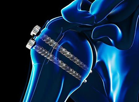
Indications for proceeding with surgical intervention include specific location of the fracture, degree of displacement, whether the fracture extends into the joint, and patient’s general physical demands. If conservative management is initiated, repeat x-rays in 7-10 days to assess any further displacement of the fracture are typically obtained. X-rays taken throughout the healing process will confirm appropriate healing of the bones. Should the fracture fail to heal, surgical treatment at a later date could be considered. This is typically an open reduction internal fixation procedure (ORIF), or a reverse replacement. Sometimes bone graft or infusion of bone morphogenic proteins (BMP) may be warranted to further stimulate fracture healing.
Conservative treatment typically includes a brief period of immobilization in a sling while refraining from active motion. As patients tolerate, their active motion can then be progressed, often with the guidance of a physical therapist. Once the fracture is healed, patients usually find they function reasonably well despite a degree of motion loss.
Proper nutrition and vitamin D and calcium supplementation can help facilitate healing. Confirming a patient’s current vitamin D level and adjusting supplementation (sometimes with a weekly mega-dose of vitamin D for 6 weeks) can help the body adequately heal the fracture. The general timeline of recovery from this injury is 3-4 months for the most dramatic improvements with continued subtle improvements over the course of the first year following injury, regardless of conservative or surgical management.